PoCUS Workshop
Program Description
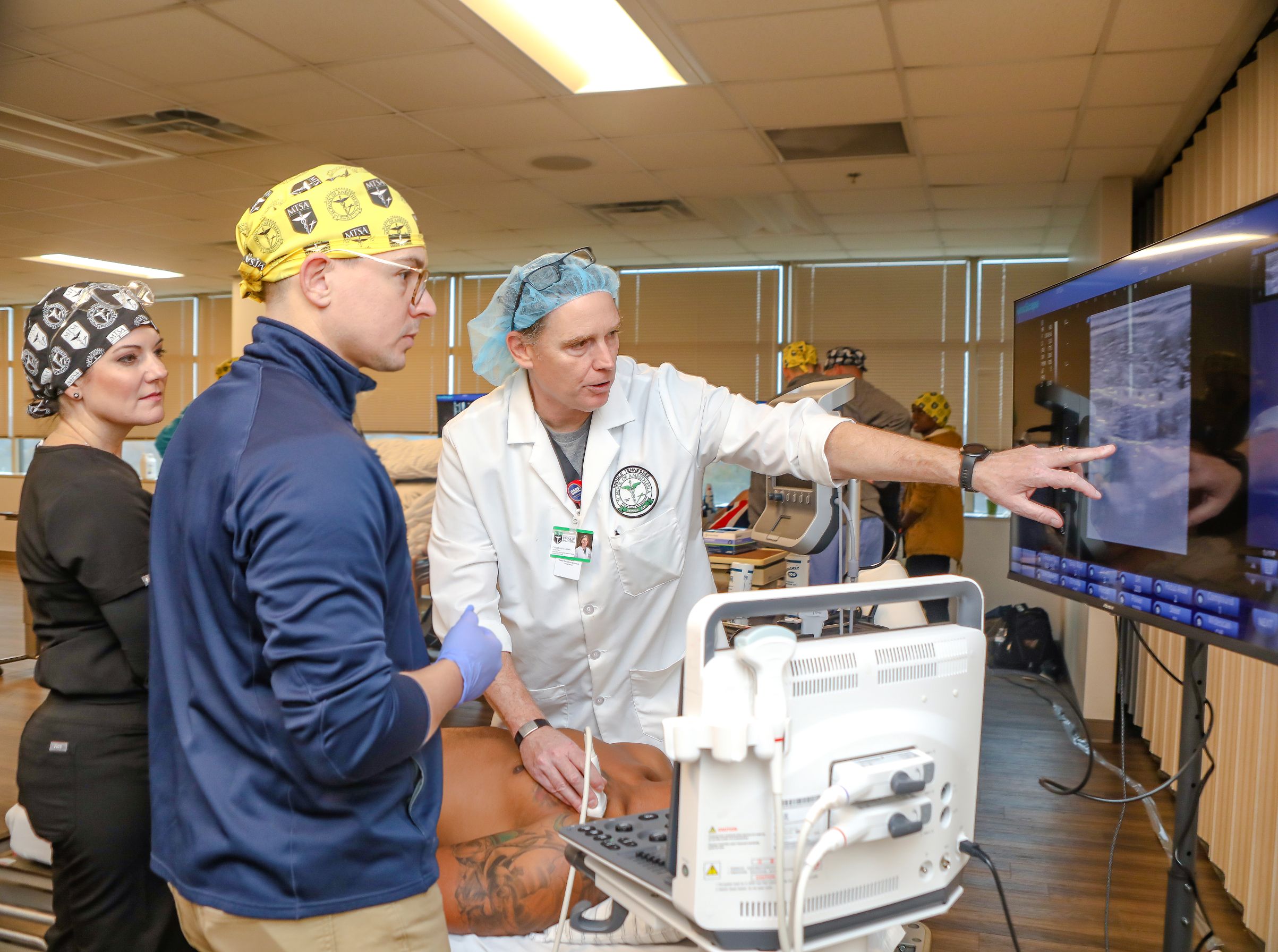
The Essentials Point-of-Care Ultrasound (PoCUS) Workshop is designed to introduce the participants to PoCUS studies that can be used as part of patient assessment in everyday clinical practice. Participants must complete the Essential PoCUS Online educational modules prior to attending the workshop.
Workshops are scheduled from 7:30 am to 12:00 pm.
Location: Middle Tennessee School of Anesthesia
Target Audience: Healthcare professionals interested in gaining knowledge and practical skills in perioperative point-of-care ultrasound.
Disclosures: Individuals involved in the planning, reviewing or execution of this activity have indicated they have no relevant financial relationships to disclose.
Questions? Contact us @ce.mtsa.edu
- Participants will need a computer and an internet connection sufficient for streaming media for the online educational modules
- Navigate to courses.mtsa.edu
- Choose the Essentials of USGRA Cadaveric Workshop you would like to attend and select "Enroll"
- Add a Voucher code if applicable and select "Recalculate"
- Select "Checkout"
- New Learner: select "Create Account"
- Already Registeres: select "Login"
- Complete the Invoice Details, Payment Options, & Important Information then select "Proceed"
- Ultrasound Principles (Physics, Sonoanatomy, Scanning Basics, Safety)
- Ultrasound Assessment of Gastric Contents
- Ultrasound-Guided Vascular Access
- Ultrasound Assessment of the Airway and Pleura
- Ultrasound Assessment of Hemodynamic Status and Intracranial Pressure
- Post-test Learner Self Evaluation
These modules must be completed prior to the hands-on workshop. The online content is self-paced and include presentations with visuals and audio.
The in-person workshop will offer you valuable practical experience through live models, high-fidelity simulations, and cadaveric specimens. It's a crucial component of your learning journey and an opportunity to apply the knowledge gained from the online modules.
The post-test and learner self-evaluation will remain locked until the learning modules are completed. All online activities must be completed prior to the hands-on workshop.
Workshops are scheduled from 7:30 am to 12:00 pm.
To receive 12 CE credits and your certificate of completion, please ensure the following:
- Completion of all online educational modules
- Achieve a minimum score of 80% on the post-test (with two attempts allowed)
- Attendance of the 4-hour hands-on workshop
- Completion of the workshop learner self-evaluation
The certificate of completion is available for download once all requirements have been met. The certificate is also emailed to the learner’s email entered in their ispringmarket account.
Module I: Ultrasound Principles (Physics, Sonoanatomy, Scanning Basics, Safety)
- Analyze the basic components of a sound wave.
- Describe how a transducer generates a sound wave.
- Compare and contrast high and low frequency ultrasound.
- Explain how an ultrasound image is created.
- Discuss the role of color Doppler in ultrasound-guided procedures.
- Relate acoustic impedance to the generation of a reflection.
- Explain the concepts of reflection, refraction, scattering, and attenuation.
- Define sonoanatomy.
- Identify different anatomic structures by their sonoanatomy.
- Review common ultrasound artifacts and their origins.
- List the essential components of an ultrasound system.
- Determine the appropriate transducer for performing various ultrasound-guided procedures.
- List the essential principles of transducer handling and manipulation.
- Relate the concepts of depth, gain, and color Doppler to ultrasound imaging.
- Define ergonomics.
- Review what is known about the bioeffects of ultrasound on cells, animals, and humans.
- Describe what is known about risk in the use of sonography and Doppler ultrasound.
- Define the ‘ALARA’ principle.
- Discuss how the provider can incorporate ALARA principles into their scanning technique to ensure safe ultrasound scanning.
Module II: Ultrasound Assessment of Gastric Contents
- Review the current perioperative fasting guidelines.
- Discuss the utility of gastric point-of-care ultrasound (POCUS) in perioperative patient management.
- State the indications for performing a gastric POCUS exam.
- Describe proper transducer placement and sonoanatomy for performing gastric POCUS.
- Qualitatively describe gastric contents based on ultrasound presentation.
- Identify parameters for determining quantitative gastric volume.
Module III: Ultrasound-Guided Vascular Access
- State the indications for vascular access in anesthetic practice.
- Review ultrasound physics and principles relative to vascular access.
- Select the optimal site for vascular access placement based on procedure and patient’s functional anatomy.
- Identify factors that place patients at increased risk for difficult IV access.
- Compare and contrast in-plane vs. out-of-plane needle insertion techniques.
- Describe essential documentation required for all vascular access procedures.
- Discuss risks and complications of ultrasound-guided vascular access procedures.
Module IV: Ultrasound Assessment of the Airway and Pleura
- Review ultrasound concepts essential to airway and pleura assessment.
- List indications for ultrasound assessment of the airway and pleura.
- Review the difficult airway algorithm.
- Describe the ‘String of Pearls’ and ‘TACA’ methods used to identify the cricothyroid membrane with ultrasound.
- List the criteria indicating a normal lung ultrasound exam.
- Explain how M-Mode ultrasound is used in lung ultrasound.
- Differentiate between the ‘Seashore Sign’ and ‘Stratosphere Sign’.
- Describe how the following pulmonary pathologies appear on ultrasound: pneumothorax, pulmonary edema, lung consolidation
- Compare and contrast a lung point and lung pulse.
Module V: Ultrasound Assessment of Hemodynamic Status and Intracranial Pressure
- Review the procedure to assess fluid volume status through ultrasound imaging of the inferior vena cava (IVC)
- Determine the fluid volume resuscitation requirements based on IVC assessment
- Demonstrate proper transducer placement and criteria for normal intracranial pressure using the optic sheath diameter.
Module I: Hands-On: Gastric Ultrasound Assessment
- Describe proper gastric ultrasound transducer placement and sonoanatomy
- Describe gastric contents qualitatively based on ultrasound presentation
- Identify parameters for determining quantitative gastric volume
Module II: Hands-On: Ultrasound-Guided Vascular Access Assessment
- Relate vascular functional anatomy to sonoanatomy
- Confirm arterial and venous flow using Doppler ultrasound
- Perform proper in-plane and out-of-plane needle insertion techniques
Module III: Hands-On: Ultrasound Assessment of the Airway and Pleura
- Identify the sonoanatomy of key anatomic structures in the anterior neck and pleura
- Demonstrate ultrasound assessment techniques to confirm endotracheal tube placement and location of the cricothyroid membrane
- Perform a lung ultrasound showing the criteria of the pleura
Module IV: Hands-On: Ultrasound Assessment of Hemodynamic Status and Intracranial Pressure
- Review the procedure to assess fluid volume status through ultrasound imaging of the inferior vena cava (IVC)
- Determine the fluid volume resuscitation requirements based on IVC assessment
- Demonstrate proper transducer placement and criteria for normal intracranial pressure using the optic sheath diameter.
Available editions:
| Product Name | Date | Location | Delivery Method | Price | |
| November 16, 2026 PoCUS Workshop | 2026-11-16 | Online | Workshop | $599.00 |